Laboratory of Tissue Engineering and Regenerative Medicine
Division of Embryology i Division of Clinical Anatomy
Faculty of Medicine
ABOUT US
Laboratory of Tissue Engineering and Regenerative Medicine was established at the Division of Embryology and the Division of Clinical Anatomy. The unit’s activities are based on the many years of experience of its staff. The scientists in the team are experts in new biologically active compounds, biomaterials, and stem cell biology, as well as epigenetics and tissue regeneration. The research work is carried out in the Division of Embryology and Clinical Anatomy in the Department of Anatomy at the MUG.
Tissue engineering is a relatively new branch of science based on biotechnology, molecular biology, nanotechnology and related sciences. The tissue engineering triad includes three main elements: cells, scaffolds and growth factors. Finding an appropriate combination of these elements enables the creation of an advanced medicinal product for a number of clinical applications. Thanks to its translational nature, tissue engineering constitutes the basis of regenerative and personalized medicine today.
Our research currently focuses on stem cells (mainly skin and adipose-derived stem cells), biologically active peptides (pro-regenerative and immunoregulatory) and epigenetic mechanisms of the regeneration and activity of the immune system. More information about the activities of the Laboratory of Tissue Engineering and Regenerative Medicine is available at www.medreg.gumed.edu.pl.


Details of some of the ongoing projects are available by clicking on the logos above.
As part of the ‘Excellence Initiative – Research University’ programme, the Laboratory also operates a core facility, offering collaborations and opportunities to perform selected scientific research on topics such as cytometric analysis, research using the Luminex platform, and 3D printing.
We cooperate with research teams from Poland and abroad:
- Departament of Surgical Oncology of the MUG
- Division of Plastic Surgery of the MUG
- Department of Dermatology, Venereology and Allergology of the MUG
- Department of Biochemistry of the MUG
- International Research Agenda, 3P – Medicine (Preventive, Personalized, Precision)
- Division of Medical Laboratory Diagnostics of the MUG
- Department of Biology and Pharmaceutical Botany of the MUG
- Department of Molecular Biotechnology and Microbiology, Gdańsk University of Technology
- Department of Polymer Technology, Gdańsk University of Technology
- Faculty of Biotechnology, the University of Wrocław
- Department of Molecular Biology, University of Gdańsk
- Department of Molecular Biotechnology, University of Gdańsk
- Department of Biomedical Chemistry, University of Gdańsk
- Department of Biomaterials, Institute of Ceramics and Building Materials, Warsaw, Łukasiewicz Research Network
- M. Nencki Institute of Experimental Biology, Polish Academy of Sciences, Warsaw
- Department of Clinical Chemistry, Lund University
- Weatherall Institute of Molecular Medicine, University of Oxford
OUR CAPACITY AND SCOPE OF RESEARCH
1. 3D Printer – the ability to create 3D structures based on gels, biomaterials, cells, creation of organoids for cytotoxicity studies, molecular studies, and optimisation of cell model creation.

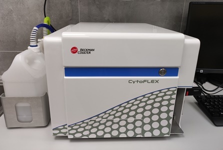
2. Flow cytometer – immunophenotypic analyses of cells, cell viability studies, nanomaterial analyses, and selected cell activity studies. More >>>
The CytoFLEX flow cytometer allows verification of multiple parameters (max. 13) in a single sample. The cytometric analysis allows the evaluation of the activity of different types of cells cultured in vitro or obtained from patients.
Examples of tests and assessments: – the percentage of dividing cells (Edu test – alternative to BrdU), – percentage undergoing apoptosis (activation of caspases and externalisation of annexin), – cell viability (PI staining, DAPI, calcein), – cell cycle (DNA content) or – determination of cell size and granularity.
Examples of tests and assessments:
- percentage of dividing cells (Edu test – alternative to BrdU),
- percentage of cells undergoing apoptosis (caspase activation and annexin externalisation),
- cell viability (PI staining, DAPI, calcein),
- cell cycle (DNA conte)
- determination of cell size and granularity.
In addition, changes under the influence of the agent/compound/applied conditions can be traced by checking for the occurrence of, among other thing
- allergic reaction (basophil activation using whole blood),
- activation of the immune system (analysis of PBMCs isolated from the patient’s blood),
- cell viability (percentage of live/dead cells).
Cytometry also allows for:
- confirmation of the immunophenotype of cells (set of surface markers characteristic of e.g. stem cells, endothelial cells, etc.),
- nanoanalysis of small molecules (e.g. exosomes).
3. Luminex – a platform allowing the analysis of multiple analytes, e.g. proteins, and cytokines, in a single sample
The method enables the analysis of multiple cytokines, growth factors, and enzymes in a single solution sample (min. 50 uL), e.g. plasma, serum, cultured medium (we study in vitro cell activity), or other fluids. The method is based on the detection of factors using fluorochrome-conjugated monoclonal antibodies. Examples of analytes assayed: IL-6, TNFα, IL-1β, IFNɣ, IL-8, IL-10, IL-4, MCP-1, Anti-SSA/Ro52, Anti-SSA/Ro60, Anti-SSB/La, Anti-C1q antibodies, C1q complement components, C3b, C2, C5a, C5, C4b TIMP inhibitors, MMP enzymes, other inflammation/sepsis markers MFI, sFasL, sFas/TNFRSF6, PAI-1 (total), sICAM.
Examples of analysis options >>>

4. Cell culture laboratory
As part of our core facility, we also offer isolation and culture of primary cells from patients (mainly from adipose tissue and skin) and immortalised cell lines. The laboratory has, among other things, laminar chambers and CO2 incubators for long-term cell cultures. It is possible to test and carry out biological analyses of new compounds, and potential drugs (regenerative, anti-cancer, immunomodulatory profile). The laboratory also provides histological evaluation of tissues (paraffin blocks, frozen tissues, histological staining).
RESEARCH TEAM
The research team includes:
- Prof. Michał Pikuła
- Milena Deptuła, Ph.D.
- Aneta Skoniecka, Ph.D.
- Agata Tymińska, M.A.
- Małgorzata Zawrzykraj, M.A.
- Katarzyna Czerwiec, M.A.
PUBLICATIONS
Many publications have been developed based on the results of the research obtained by using the knowledge, experience of scientists from the Laboratory and its equipment, including.
1. Jedrzejewska A, Kawecka A, Braczko A, Romanowska-Kocejko M, Stawarska K, Deptuła M, Zawrzykraj M, Franczak M, Krol O, Harasim G, Walczak I, Pikuła M, Hellmann M, Kutryb-Zając B. Changes in Adenosine Deaminase Activity and Endothelial Dysfunction after Mild Coronavirus Disease-2019 Int J Mol Sci. 2023 Aug 24;24(17):13140 doi: 10.3390/ijms241713140
2. Milena Deptuła, Małgorzata Zawrzykraj, Justyna Sawicka, Adrianna Banach-Kopeć, Robert Tylingo, Michał Pikuła Application of 3D- printed hydrogels in wound healing and regenerative medicine Biomed Pharmacother. 2023 Nov:167:115416. doi: 10.1016/j.biopha.2023.115416
3. Dorota Piechota, Patrycja Wardaszka, Wioletta Barańska-Rybak 1, Maciej Jaœkiewicz, Wojciech Kamysz, Milena Deptuła, Michał Pikuła, Ryszard Ostaszewski Interdisciplinary approach to synthesis, stability and antimicrobial activity of silver nanoparticles Postepy Dermatol Alergol. 2023 Jun;40(3):390-397. doi: 10.5114/ada.2023.128978
4. Adrian Kobiela, Lilit Hovhannisyan, Paulina Jurkowska, Jorge Bernardino de la Serna, Aleksandra Bogucka, Milena Deptuła, Argho Aninda Paul, Kinga Panek, Ewa Czechowska, Michał Rychłowski, Aleksandra Królicka, Jacek Zieliński, Susanne Gabrielsson, Michał Pikuła, Magdalena Trzeciak, Graham S Ogg, Danuta Gutowska-Owsiak Excess filaggrin in keratinocytes is removed by extracellular vesicles to prevent premature death and this mechanism can be hijacked by Staphylococcus aureus in a TLR2-dependent fashion Extracell Vesicles 2023 Jun;12(6):e12335. doi: 10.1002/jev2.12335. Więcej >>>>
5. Dzierżyńska M., Sawicka J., Deptuła M., Sosnowski P., Sass P., Peplińska B., Pietralik-Molińska Z., Fularczyk M., Kasprzykowski F., Zieliński J., Kozak M., Sachadyn P., Pikuła M., Rodziewicz-Motowidło S. Release systems based on self-assembling RADA16-I hydrogels with a signal sequence which improves wound healing processes. Sci Rep. 2023 Apr 18;13(1):6273. doi: 10.1038/s41598-023-33464-w. PMID: 37072464.
6. Katarzyna Czerwiec, Małgorzata Zawrzykraj, Milena Deptuła, Aneta Skoniecka, Agata Tymińska, Jacek Zieliński, Adam Kosiński, Michał Pikuła Adipose-Derived Mesenchymal Stromal Cells in Basic Research and Clinical Applications. Int J Mol Sci 2023 Feb 15; 24(4):3888. doi: 10.3390/ijms24043888.
1. Katarzyna Kuncewicz, Claire Battin, Katarzyna Węgrzyn, Adam Sieradzan, Anna Wardowska, Emilia Sikorska, Irma Giedrojć, Pamela Smardz, Michał Pikuła, Peter Steinberger, Sylwia Rodziewicz-Motowidło, Marta Spodzieja, Targeting the HVEM protein using a fragment of glycoprotein D to inhibit formation of the BTLA/HVEM complex. Bioorg Chem. 2022 May;122:105748. doi: 10.1016/j.bioorg.2022.105748. Epub 2022 Mar 19.
2. Paweł Sosnowski, Piotr Sass, Paulina Słonimska, Rafał Płatek, Jolanta Kamińska, Jakub Baczyński Keller, Piotr Mucha, Grażyna Peszyńska-Sularz, Artur Czupryn, Michał Pikuła, Arkadiusz Piotrowski, Łukasz Janus, Sylwia Rodziewicz-Motowidło, Piotr Skowron, Paweł Sachadyn, Regenerative Drug Discovery Using Ear Pinna Punch Wound Model in Mice. Pharmaceuticals (Basel). 2022 May 16;15(5):610. doi: 10.3390/ph15050610.
3. Adrian Kobiela, Joanna E Frackowiak, Anna Biernacka, Lilit Hovhannisyan, Aleksandra E Bogucka, Kinga Panek, Argho Aninda Paul, Joanna Lukomska, Xinwen Wang, Eleni Giannoulatou, Aleksandra Krolicka, Jacek Zielinski, Milena Deptula, Michal Pikula, Susanne Gabrielsson, Graham S Ogg, Danuta Gutowska-Owsiak, Exposure of Keratinocytes to Candida Albicans in the Context of Atopic Milieu Induces Changes in the Surface Glycosylation Pattern of Small Extracellular Vesicles to Enhance Their Propensity to Interact With Inhibitory Siglec Receptors. Front Immunol. 2022 Jun 9;13:884530. doi: 10.3389/fimmu.2022.884530. eCollection 2022.
1. Kosikowska-Adamus P., Sikorska E., Wyrzykowski D., Walewska A., Golda A., Deptuła M., Obuchowski M., Prahl A., Pikuła M., Lesner A. 2021. Lipidation of Temporin-1CEb Derivatives as a Tool for Activity Improvement, Pros and Cons of the Approach. Int. J. Mol. Sci. 22, no. 13: 6679. doi.org/10.3390/ijms22136679.
2. Deptuła M., Brzezicka A., Skoniecka A., Zieliński J., Pikuła M., Adipose-derived stromal cells for nonhealing wounds: Emerging opportunities and challenges Medicinal Research Reviews 2021 Jul; 41(4):2130-2171 doi: 10.1002/med.21789 Więcej >>>>
3. Sawicka J., Iłowska E., Deptuła M., Sosnowski P., Sass P., Czerwiec K., Chmielewska K., Szymańska A., Pietralik-Molińska Z., Kozak M., Sachadyn P., Pikuła M., Rodziewicz-Motowidło S., Functionalized Peptide Fibrils as a Scaffold for Active Substances in Wound Healing. Int J Mol Sci. 2021; 22(8): 3818. doi: 10.3390/ijms22083818.
4. Mazuryk J., Puchalska I., Koziński K., Ślusarz M.J., Ruczyński J., Rekowski P., Rogujski P., Płatek R., Wiśniewska M.B., Piotrowski A., Janus Ł., Skowron P.M., Pikuła M., Sachadyn P., Rodziewicz-Motowidło S., Czupryn A., Mucha P. PTD4 Peptide Increases Neural Viability in an In Vitro Model of Acute Ischemic Stroke. Int J Mol Sci. 2021; 22(11):6086.
5. Skoniecka A., Cichorek M., Tyminska A., Pelikant-Malecka I., Dziewiątkowski J., Melanization as unfavorable factor in amelanotic melanoma cell biology Protoplasma 2021 Jan 27.
6. Cichorek M, Kowiański P, Lietzau G, Lasek J, Moryś J. 2021. Neuroglia – development and role in physiological and pathophysiological processes Folia Morphol (Warsz). 2021;80(4):766-775. 10.5603/FM.a2021.0109.
1. Małuch I., Stachurski O., Kosikowska-Adamus P., Makowska M., Bauer M., Wyrzykowski D., Hać A., Kamysz W., Deptuła M., Pikuła M., Sikorska E., Double-Headed Cationic Lipopeptides: An Emerging Class of Antimicrobials. Int. J. Mol. Sci. 2020, 21, 8944.
2. Kuncewicz K., Battin C., Sieradzan A., Karczyńska A., Orlikowska M., Wardowska A., Pikuła M., Steinberger P., Rodziewicz-Motowidło S., Spodzieja M., Fragments of gD Protein as Inhibitors of BTLA/HVEM Complex Formation – Design, Synthesis, and Cellular Studies. Int. J. Mol. Sci. 2020, 21, 8876.
3. Cichorek M., Ronowska A., Dzierzbicka K., Gensicka-Kowalewska M., Deptuła M., Pelikant-Maleckade I., Chloroacridine derivatives as potential anticancer agents which may act as tricarboxylic acid cycle enzyme inhibitors. Biomedicine & Pharmacotherapy Vol. 130, October 2020, 110515
4. Golab K., Krzystyniak A., Langa P., Pikuła M., Kunovac S, Borek P., Trzonkowski P., Millis J.M., Fung J., Witkowski P., Effect of serum on SmartFlare™ RNA Probes uptake and detection in cultured human cells. Biomed J Sci Tech Res. 2020; 28(4): 21788-21793.
5. Biernat M., Ciołek L., Dzierżyńska M., Oziębło A., Sawicka J., Deptuła M., Bauer M., Kamysz W., Pikuła M., Jaegermann Z., Rodziewicz‐Motowidło S., Porous chitosan/ZnO‐doped bioglass composites as carriers of bioactive peptides. Int J Appl Ceram Technol. 2020;17: 2807-2816.
6. Wardowska A., Smoleńska Ż., Lisowska K.A., Zdrojewski Z., Pikuła M., Dendritic cells’ characteristics in patients with treated systemic lupus erythematosus. Acta Biochim Pol. 2020 Jul 30;67(3): 417-429. doi: 10.18388/abp.2020_5416.
7. Sawicka J., Dzierżyńska M., Wardowska A., Deptuła M., Rogujski P., Sosnowski P., Filipowicz N., Mieczkowska A., Sass P., Pawlik A., Hać A., Schumacher A., Gucwa M., Karska, N., Kamińska J., Płatek R., Mazuryk J., Zieliński J., Kondej K., Młynarz P., Mucha P., Skowron P., Janus Ł., Herman-Antosiewicz A., Sachadyn P., Czupryn A., Piotrowski A., Pikuła M., Rodziewicz-Motowidło S., Imunofan—RDKVYR Peptide—Stimulates Skin Cell Proliferation and Promotes Tissue Repair. Molecules 2020, 25, 2884.
8. Wardowska A., Komorniczak M., Skoniecka A., Bułło-Piontecka B., Lisowska K.A., Dębska-Ślizień M.A., Pikuła M., Alterations in peripheral blood B cells in systemic lupus erythematosus patients with renal insufficiency. Int Immunopharmacol. 2020 Apr 1;83:106451.
9. Siebert A., Deptuła M., Cichorek M., Ronowska A., Cholewiński G., Rachon J., Anticancer Properties of Amino Acid and Peptide Derivatives of Mycophenolic Acid. Anti-Cancer Agents in Med. Chem. 2020 May
10. Wardowska A. The epigenetic face of lupus: Focus on antigen-presenting cells Int Immunopharmacol 2020 Apr;81:106262;
11. Skowron P.M., Krawczun N., Zebrowska J., Krefft D., Zolnierkiewicz O., Bielawa M., Jezewska-Frackowiak J., Janus L., Witkowska M., Palczewska M., Schumacher A., Wardowska A., Deptula M., Czupryn A., Mucha P., Piotrowski P., Sachadyn P., Rodziewicz-Motowidlo S., Pikula M., Zylicz-Stachula A. A vector-enzymatic DNA fragment amplification-expression technology for construction of artificial, concatemeric DNA, RNA and proteins for novel biomaterials, biomedical and industrial applications. Materials Science and Engineering: C. Vol. 108, March 2020, 110426
12. Skowron P.M., Krawczun N., Zebrowska J., Krefft D., Zolnierkiewicz O., Bielawa M., Jezewska-Frackowiak J., Janus L., Witkowska M., Palczewska M., Schumacher A., Wardowska A., Deptula M., Czupryn A., Mucha P., Piotrowski P., Sachadyn P., Rodziewicz-Motowidlo S., Pikuła M., Zylicz-Stachula A., Data regarding a new, vector-enzymatic DNA fragment amplification-expression technology for the construction of artificial, concatemeric DNA, RNA and proteins as well as biological effects of selected polypeptides obtained this method. Data in Brief, 2019 Dec 31;28:105069. doi: 10.1016/j.dib.2019.105069. eCollection 2020 Feb.
13. Zieliński J., Jaworski R., Pikuła M., Jaśkiewicz J., Girnyi S., Pałubicka A., But M. The Influence of Pre-Operative Chemo- and Radiotherapy in Patients with Colon Cancer on Wound Healing in the Material from a Single Center. European Journal of Surgical Oncology, Feb 2020, vol. 46, nr 2, s. e109
14. Deptuła M., Karpowicz P., Wardowska A., Sass P., Sosnowski P., Mieczkowska A., Filipowicz N., Dzierżyńska M., Sawicka J., Nowicka E., Langa P., Schumacher A., Cichorek M., Zieliński J., Kondej K., Kasprzykowski F., Czupryn A., Janus Ł., Mucha P., Skowron P., Piotrowski A., Sachadyn P., Rodziewicz-Motowidło S., Pikuła M. , Development of a Peptide Derived from Platelet-Derived Growth Factor (PDGF-BB) into a Potential Drug Candidate for the Treatment of Wounds. Advances in Wound Care. 2020 Dec;9(12): 657-675. doi: 10.1089/wound.2019.1051.
1. Sass P. , Sosnowski P., Podolak-Popinigis J., Górnikiewicz B., Kamińska J., Deptuła M., Nowicka E., Wardowska A., Ruczyński J., Rekowski P., Rogujski P., Filipowicz N., Mieczkowska A., Peszyńska-Sularz G., Janus Ł., Skowron P., Czupryn A., Mucha P., Piotrowski A., Rodziewicz-Motowidło S., Pikuła M., Sachadyn P. Epigenetic inhibitor zebularine activates ear pinna wound closure in the mouse. EBioMedicine. 2019; 46: 317-329 Więcej >>>>
2. Wardowska A., Komorniczak M., Bułło-Piontecka B., Dębska-Ślizień A., Pikuła M. Transcriptomic and epigenetic alterations in dendritic cells correspond with chronic kidney disease in lupus nephritis. Front. Immunol. 2019; 10: 2026. doi: 10.3389/fimmu.2019.02026
3. Palubicka A., Jaworski R., Wekwejt M., Swieczko-Zurek B., Pikula M., Jaskiewicz J., Zielinski J. Surgical Site Infection after Breast Surgery: A Retrospective Analysis of 5-Year Postoperative Data from a Single Center in Poland. Medicina. 2019; 55(9): 512.
4. Specjalski K., Maciejewska E., Pawłowski R., Zieliński M., Trzonkowski P., Pikuła M., Jassem E. Changing microRNA Expression during Three-Month Wasp Venom Immunotherapy. Immunol Invest. 2019; 24: 1-9.
5. Deptuła M., Zieliński J., Wardowska A., Pikuła M. Wound healing complications in oncological patients: perspectives for cellular therapy. Adv Dermatol Allergol. 2019; 36(2): 139-146.
6. Girnyi S., Serkies K., Jaworski R., But M., Wiski A., Komornicka J., Pikuła M., Zieliński J. Skin giant neoplastic ulcers – treatment options and clinical challenges. Nowotwory. Journal of Oncology 2019; 69(1): 12-17.
7. Krzystyniak A., Baczynska E., Magnowska M., Antoniuk S., Roszkowska M., Zareba-Koziol M., Das N., Basu S., Pikuła M., Wlodarczyk J. Prophylactic Ketamine Treatment Promotes Resilience to Chronic Stress and Accelerates Recovery: Correlation with Changes in Synaptic Plasticity in the CA3 Subregion of the Hippocampus. Int J Mol Sci. 2019; 8; 20(7)
8. Kamińska J., Kamińska J., Langa P., Deptuła M., Zieliński J., Sachadyn P., Wardowska A., Pikuła M. Transcriptional activity of epigenetic remodelling genes declines in keratinocytes after in vitro Expansion. Adv Med Sci. 2019; 64: 274-279.
1. Brzezicka A., Kondej K., Błażyńska-Spychalska A., Spychalski P., Jankau J., Pikuła M. Zastosowanie komórek macierzystych tkanki tłuszczowej w medycynie – najnowsze trendy. Chirurgia Plastyczna i Oparzenia. 2018; 6(3): 79-85.
2. Gójska-Grymajło A., Zieliński M., Wardowska A., Gąsecki D., Pikuła M., Karaszewski B. CXCR7+ and CXCR4+ stem cells and neuron specific enolase in acute ischemic stroke patients. Neurochem Int. 2018; 120: 134-139
3. Mieczkowska A., Schumacher A., Filipowicz N., Wardowska A., Zieliński M., Madanecki P., Nowicka E., Langa P., Deptuła M., Zieliński J., Kondej K., Renkielska A., Buckley P.G., Crossman D.K., Crowley M.R., Czupryn A., Mucha P., Sachadyn P., Janus Ł., Skowron P., Rodziewicz-Motowidło S., Cichorek M., Pikuła M., Piotrowski A. Immunophenotyping and transcriptional profiling of in vitro cultured human adipose tissue derived stem cells. Sci Rep. 2018; 27,8(1):11339 Więcej >>>>
4. Schumacher A., Cichorek M., Pikuła M. Komórki macierzyste tkanki tłuszczowej w inżynierii tkankowej i terapii trudno gojących się ran [Adipose-derived stem cells for tissue engineering and therapy of non-healing wounds]. Postepy Hig i Med. Dośw. 2018; 72: 806-821
5. Deptuła M., Wardowska A., Dzierżyńska M., Rodziewicz-Motowidło S., Pikuła M. Antibacterial Peptides in Dermatology–Strategies for Evaluation of Allergic Potential. Molecules. 2018; 23(2), 414; doi:10.3390/molecules23020414
36. Mucha P., Pieszko M., Miszka A., Ruczyński J., Rekowski P., Załuska I., Kozłowska A., Schumacher A., Deptuła M., Pikuła M. Ru(II)-mediated synthesis and bioactivity evaluation of 1,4,5-trisubstituted N-phthalimido protected 5-bromo-1,2,3-triazolic amino acid. Lett Org Chem. 2018; 15(4): 282-289.
PRICE LIST
The laboratory will perform analyses according to individual orders with adaptation to specific needs of contractors. The cost of the service is priced individually for external customers.
CONTACT

Prof. Michał Pikuła
Laboratory of Tissue Engineering and Regenerative Medicine
Department of Anatomy and Division of Embryology
Medical University of Gdańsk
Dębinki 1 St.
80-211 Gdańsk
Phone 58 349 13 68
Mail michal.pikula@gumed.edu.pl
www www.medreg.gumed.edu.pl


photo Paweł Sudara/MUG and private archive