Laboratory of Fluorescence and Confocal Microscopy
Division of Anatomy and Neurobiology
Faculty of Medicine
ABOUT US
Laboratory of Fluorescence and Confocal Microscopy was established under the “Excellence Initiative – Research University” Program as a core facility laboratory at the Division of Anatomy and Neurobiology of the MUG. The Laboratory’s work involves outstanding specialists in fluorescence microscopy and confocal microscopy. The Laboratory’s research team has set as its goal the dissemination of advanced microscopic imaging, and thus supports staff and doctoral students in deepening their knowledge of microscopic techniques by providing access to specialized equipment.
OUR POTENTIAL
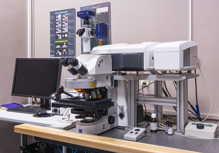
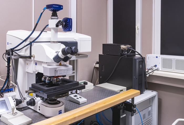
1. Zeiss LSM 880 confocal microscopy system with AiryScan super-resolution attachment interfacing with Zeiss Axio Imager.Z2 microscope
The system allows the analysis of preparations stained by histochemical and immunohistochemical methods (frozen sections, fixed cell cultures) prepared on basic slides of 25mm x 75mm size by a system based on a straight microscope that allows brightfield operation, using Nomarski contrast (DIC).
2. Zeiss AxioScan.Z1 histology slide scanner
Integrated system for automatic scanning of preparations stained by histochemical or immunohistochemical methods allows the use of different types of magazines for slides that can hold: up to 100 slides measuring 25 mm x 75 mm, up to 2 slides measuring 50mm x 75mm and 1 slide measuring 100mm x 75mm.
3. PALM Microbeam laser microdissection system working with AxioObserver microscope 7 Zeiss
System based on an inverted microscope with motorized objective turret holder with automatic recognition of installed lenses.
4. Olympus MVX 10 Olympus monoscopic microscope
Allows analysis of preparations stained by histochemical and immunohistochemical methods (frozen sections, fixed cell cultures) prepared on basic slides measuring 25mm x 75mm by a system based on a monoscopic microscope adapted to macroscopic object imaging (without the need for image assembly).
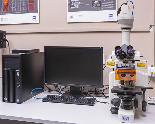
5. Nikon Eclipse E600 microscope with Nikon DS-Ri2 camera, Nikon, Japan
Allows analysis of preparations stained by histochemical and immunohistochemical methods (frozen sections, fixed cell cultures) prepared on basic slides of 25mm x 75mm size by a system based on a simple fluorescence microscope.
SCOPE OF SERVICES
– morphological evaluation of biological specimens based on light microscopy methods, fluorescence microscopy and confocal microscopy,
– qualitative evaluation of preparations labeled by histochemical and immunocytochemical methods in single and multiple stains – with special emphasis on the cellular localization of the tested compounds,
– mapping of the tested compounds within the studied structures – analysis of the stained histochemical and immunocytochemical methods in staining single and multiple serial sections,
– quantitative analysis of selected morphological parameters using the methods of confocal microscopy,
– laser microdissection of fragments of frozen, paraffin-embedded preparations, single cells and cell fragments,
– cutting fresh and fixed tissue directly from slides,
– digital camera image acquisition, image file processing,
– recording of images in multiple formats, i.e. TIFF, JPG,
– possible editing to allow automation of processes,
– ability to create reports that include images and analysis results, creating custom report forms.
RESEARCH TEAM
The research team includes:
- Dr. Habil. Przemysław Kowiański, Assoc. Prof.
- Dr. Habil. Beata Ludkiewicz
- Dr. Habil. Sławomir Wójcik
- Aleksandra Rutkowska, Ph.D.
- Jan Henryk Spodnik, Ph.D.
- Grażyna Lietzau, Ph.D.
- Ewelina Czuba-Pakuła, M.A.
- Adriana Pszczolińska, M.A.
- Natalia Melka, M.A.
PUBLICATIONS
Publications that have been developed by using the research methods described:
1. Steliga A, Lietzau G, Wójcik S, Kowiański P. Transient cerebral ischemia induces the neuroglial proliferative activity and the potential to redirect neuroglial differentiation. J Chem Neuroanat. 2022 Nov 17;127:102192. doi: 10.1016/j.jchemneu.2022.102192
2. Piekarska K, Urban-Wójciuk Z, Kurkowiak M, Pelikant-Małecka I, Schumacher A, Sakowska J, Spodnik J, Arcimowicz Ł, Zielińska H, Tymoniuk B, Renkielska A, Siebert J, Słomińska E, Trzonkowski P, Hupp T, Marek-Trzonkowska N. Mesenchymal stem cells transfer mitochondria to allogeneic Tregs in an HLA-dependent manner improving their immunosuppressive activity. Nat. Commun. 2022 : vol. 13, art. ID 856, s. 1-20
3. Velasco-Estevez M, Koch N, Klejbor I, Caratis F, Rutkowska A. Mechanoreceptor Piezo1 is downregulated in multiple sclerosis brain and is involved in the maturation and migration of oligodendrocytes in vitro. Front. Cell. Neurosci. 2022 : vol. 16, art. ID 914985, s. 1-9
4. Velasco-Estevez M, Koch N, Klejbor I, Laurent S, Dev Kumlesh K., Szutowicz A, Sailer A.W., Rutkowska A. EBI2 is temporarily upregulated in MO3.13 oligodendrocytes during maturation and regulates remyelination in the organotypic cerebellar slice model. Int. J. Mol. Sci. 2021 : vol. 22, nr 9, art. ID 4342, s. 1-15
5. A. Ebertowska, B.Ludkiewicz, N. Melka, I. Klejbor J. Moryś. The influence of early postnatal chronic mild stress stimulation on the activation of amygdala in adult rat J Chem Neuroanat 2020;104:10174
6. Nicotine-induced CREB and DeltaFosB activity is modified by caffeine in the brain reward system of the rat. Kowiański P, Lietzau G, Steliga A, Czuba E, Ludkiewicz B, Waśkow M, Spodnik JH, Moryś J. J Chem Neuroanat. 2018 Mar;88:1-12. doi: 10.1016/j.jchemneu.2017.10.005. Epub 2017 Nov 1
7. J Sidor-Kaczmarek, M Cichorek, JH Spodnik, S Wójcik, J Moryś. Proteasome inhibitors against amelanotic melanoma. Cell Biol Toxicol. 2017; 33(6): 557–573. Published online 2017 Mar 9. doi: 10.1007/s10565-017-9390-0
8. S. Wójcik, JH Spodnik, J Dziewiątkowski, E Spodnik, J Moryś. Morphological Changes within the Rat Lateral Ventricle after the Administration of Proteasome Inhibitors. PLoS One. 2015; 10(10): e0140536. Published online 2015 Oct 19. doi: 10.1371/journal.pone.0140536
9. Wójcik, S., Spodnik, J.H., Spodnik, E., Dziewiątkowski, J., Moryś, J. Nigrostriatal pathway degeneration in rats after intraperitoneal administration of proteasome inhibitor MG-132. 2014. Folia Neuropathologica 52(1):41-55. DOI: 10.5114/fn.2014.41743
10. Steliga A, Waśkow M, Karwacki Z, Wójcik S, Lietzau G, Klejbor I, Kowianski P. Transcription factor Pax6 is expressed by astroglia after transient brain ischemia in the rat model. Folia Neuropathol. 2013;51(3):203-13. doi:10.5114/fn.2013.37704. PMID: 24114637
PRICE LIST
The laboratory performs research and analysis according to individual projects. Service costs are valued individually.
CONTACT

Dr. Habil. Sławomir Wójcik
Laboratory of Fluorescence and Confocal Microscopy
Division of Anatomy and Neurobiology
Medical University of Gdańsk
ul. Dębinki 1
80-211 Gdańsk
Phone: 58 349 14 02
E-Mail slawomir.wojcik@gumed.edu.pl
photo Paweł Sudara/MUG


