Laboratory of Imaging Pathology of Cell Structure and Function
Medical University of Gdańsk
As part of the “Excellence Initiative – Research University” program, the core facility – the Laboratory of Imaging Pathology of Cell Structure and Function – is being expanded at GUMed. The unit was established in 2012, thanks to the experience of employees at the Department of Physiopathology and Division of Pathology and Experimental Rheumatology operating as a part of the Faculty of Medicine at the Medical University of Gdansk. The laboratory cooperates with numerous research and clinical units of the Medical University of Gdansk, as well as with the University of Gdansk and Gdansk University of Technology.
In addition to advanced cellular analyses and molecular mechanisms of the involvement of lymphocytes, and other cells in the development and progression of autoimmune diseases, it also offers analogous studies of human aging processes and the development of diseases associated with old age.
Our potential
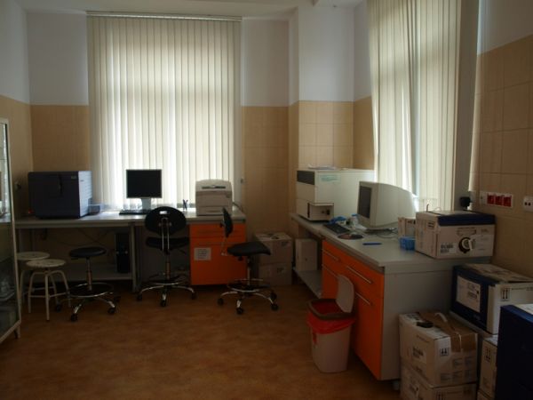
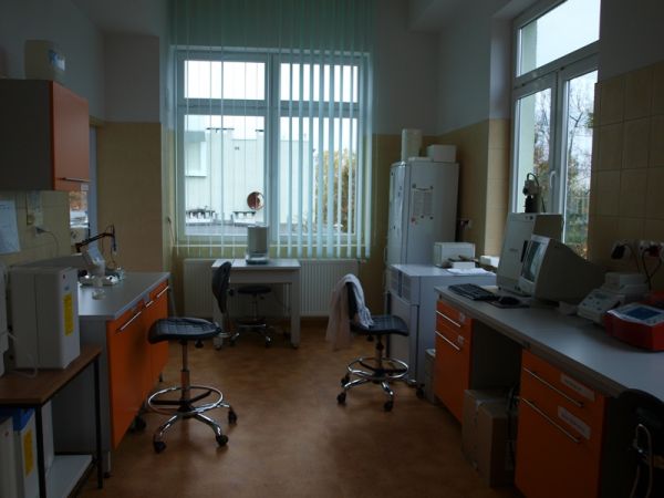
Laboratory of Flow Cytometry and Cell Imaging
Multiparameter assessment of immune and other cell properties and activities:
Evaluation of phenotypes:
Surface staining of phenotypes (markers on cells and intracellular antigens) by multiparameter flow cytometry technique – working with freshly isolated blood lymphocytes and monocytes, as well as isolated from lymphoid organs (thymus, lymph nodes), cultured cells of various origins (including cell lines).
Functional tests of:
- lymphocytes:
- proliferation capacity (a proprietary, quantitative cytometric method (Dividing Cell Tracking™) that determines cycle length, number of generations, and other dynamic parameters of proliferation in cells of known phenotype, requiring no population separation or radioisotopes; cell cycle assessment by DNA quantity assay),
- susceptibility to apoptosis and necrosis – measurement of changes in mitochondrial potential, assessment of surface expression of phosphatidylserine, assessment of DNA dye penetration
- lyintracellular T lymphocyte cytotoxicity and caspase activity tests, in living cells,
- semiquantitative assessment of phosphorylation of selected signaling proteins using the Phos-FLOW technique),
- semiquantitative evaluation of calcium signal
- semiquantitative evaluation of intracellular calpain protease activity
- semiquantitative assessment of intracellular cytokine production
- monocytes, granulocytes, macrophages:
- measuring phagocytosis capacity and oxygen burst
- semiquantitative assessment of intracellular cytokine production
Sorting phenotypically pure cell populations (lymphocytes, monocytes, etc.)
Examination of secreted cytokines and other biologically active proteins in body fluids using the CBA method (quantitative flow cytometry), enabling simultaneous measurement of up to 6 factors in 50 µL of specimen (material and cost savings as compared with the ELISA technique, while maintaining measurement accuracy and evaluation of absolute concentration).
Equipment: flow cytometers: FACScan™, FACSVerse™, a FACSAria III™ cytometer-sorter (Becton Dickinson), a Nikon Eclipse high-performance universal fluorescence microscope, and IN Cell Analyzer™ 2200/2000is automated microscope (GE Healthcare Life Sciences) enabling rapid, automated imaging and analysis of live cells (up to 7 days of observation) and fixed cells, purchased with a grant from the Ministry of Science and Higher Education for the GUMed and UG Network of Pathology Imaging Laboratories for Cell Structure and Function.
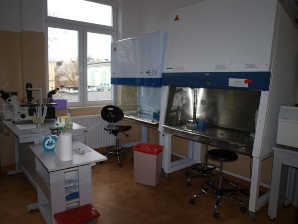
Tissue Culture Laboratory
In vitro cell cultures – sterile work, performing short-term and long-term cell cultures to study the effects of specific factors on cell proliferation or apoptosis.
Equipment: laminar flow chambers (Biosafety grade) and CO2 and O2 incubators (with the ability to conduct cell cultures in physiological, lower than atmospheric, oxygen concentrations).
Laboratory of Molecular Biology
Molecular techniques used:
RT-PCR, Western-Blot, real-time PCR (quantitative). Full equipment for this type of lab, including:
- laminar flow chamber,
- thermocyclers (including one adapted to perform PCR reactions in material on microscope slides),
- centrifuges,
- ultra-pure water system,
- epoch spectrophotometer,
- computerized archiving of test results.
Our team
The research team includes experienced scientists:
- Prof. Jacek M. Witkowski
- Prof. Ewa Bryl
- Jacek Brożek, Ph.D.
- Agnieszka Daca, Ph.D.
- Dr Habil. Katarzyna Lisowska
- Katarzyna Ruckemann-Dziurdzińska, Ph.D.
- Monika Soroczyńska-Cybula, Ph.D.
- Dr. Habil. Anna Wardowska
- Klaudia Ciesielska-Figlon, M.A.
- Natalia Frankowska, M.A. – coordinator for contact with persons interested in cooperation/using the services of the laboratory
- Paulina Łopatniuk, M.D.
- Agnieszka Odachowska, M.A. – office
doctoral students:
- Marcin Kutek, M.D.
- Mateusz Maziewski, M.A.
- Paweł Mikłaszewicz, M.D.
- Małgorzata Pindel, M.A.
- Adam Plotka, M.D.
- Jakub Ruszkowski, M.D.
- Łukasz Szałach, M.D.
Price list:
The laboratory performs research and analysis according to individual projects. The cost of the service is priced individually.
Some of the publications that were based on the research results obtained through the use of the research apparatus at hand:
1. The methodological quality and clinical applicability of meta-analyses on probiotics in 2020: a cross-sectional study
RUSZKOWSKI JAKUB, Majkutewicz Katarzyna, RYBKA EWELINA, KUTEK MARCIN, DĘBSKA-ŚLIZIEŃ ALICJA, WITKOWSKI JACEK MACIEJ. – Biomed. Pharmacother. – 2021 : vol. 142, art. ID 112044, s. 1-10, bibliogr. 49 items., abstracts. English.
2. The influence of biologics on the microbiome in immune-mediated inflammatory diseases: a systematic review
RUSZKOWSKI JAKUB, DACA AGNIESZKA, SZEWCZYK ADRIAN, DĘBSKA-ŚLIZIEŃ ALICJA, WITKOWSKI JACEK M.. – Biomed. Pharmacother. – 2021 : vol. 141, art. ID 111904, s. 1-19, bibliogr. 35 items., v
Gut microbiota in depression : a focus on ketamine – WILKOWSKA ALINA, SZAŁACH ŁUKASZ PIOTR, CUBAŁA WIESŁAW JERZY. – Front. Behav. Neurosci. – 2021 : vol. 15, art. ID 693362, s. 1-12, bibliogr. items., abstracts. English.
3. Nigella sativa oil inhibits proliferation and stimulates apoptosis of human lymphocytes in vitro
CIESIELSKA-FIGLON KLAUDIA, LISOWSKA KATARZYNA A., Mikosik-Roczynska Anna, WITKOWSKI JACEK M.. – Hum. Immunol. – 2021 : vol. 82, nr 8, s. 608-614, bibliogr. 16 items., abstracts. English.
4. Effect of choline chloride based natural deep eutectic solvents on aqueous solubility and thermodynamic properties of acetaminophen
Warmińska Dorota, Nowosielski Bartosz, SZEWCZYK ADRIAN, RUSZKOWSKI JAKUB, PROKOPOWICZ MAGDALENA. – J. Mol. Liq. – 2021 : vol. 323, art. 114834, s. 1-15, bibliogr. 59 items., abstracts. English.
5. Targeting impaired antimicrobial immunity in the brain for the treatment of Alzheimer’s disease
Fulop Tamas, Tripathi Shreyansh, Rodrigues Serafim, Desroches Mathieu, Bunt Ton, Eiser Arnold, Bernier Francois, Beauregard Pascale B, Barron Annelise E, Khalil Abdelouahed, PLOTKA ADAM, Hirokawa Katsuiku, Larbi Anis, Bocti Christian, Laurent Benoit, Frost Eric H, WITKOWSKI JACEK M.. – Neuropsychiatr. Dis. Treat. – 2021 : vol. 17, s. 1311-1339, bibliogr. 381 items., abstracts. English.
6. Associations between symptoms of constipation and sleep quality in patients with nondialysis chronic kidney disease: a cross sectional study
RUSZKOWSKI JAKUB, HELENIAK ZBIGNIEW, KRÓL EWA, TARASEWICZ AGNIESZKA, WITKOWSKI JACEK M., DĘBSKA-ŚLIZIEŃ ALICJA. – Pol. Arch. Med. Wewn. – 2021 : vol. 131, nr 6, s.512-519, bibliogr. 42 items., abstracts. English.

CONTACT
Natalia Frankowska
Department of Physiopathology
Medical University of Gdańsk
Phone: 58 349 15 10
natalia.frankowska@gumed.edu.pl