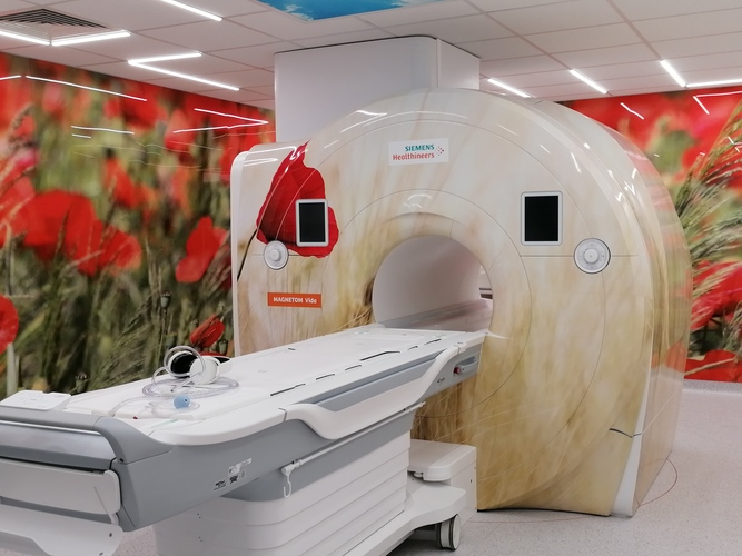
Magnetic Resonance Imaging Laboratory
Medical University of Gdańsk
The Magnetic Resonance Imaging Laboratory provides infrastructure and support to facilitate research and educational activities using magnetic resonance technology. Advanced equipment is the basis for conducting interdisciplinary research. Modern imaging methods are becoming the basis not only for clinical diagnosis, but also for progress in the field of basic science and development of new therapeutic methods. The MRI scanner at the MUG creates a collaborative space not only for researchers, but also for those conducting clinical research, as well as healthcare-related manufacturing companies.
The state-of-the-art 3T Siemens Magnetom Vida scanner allows for the development of innovative products and technologies that enable the advancement of diagnostics and therapy of civilization diseases, as well as the development of new ways of diagnostics and therapy or its monitoring.

The laboratory is equipped with MRI 3T Magnetom Vida (Siemens Healthineers, Erlangen, Germany). We have the following coils to choose from:
- 64-channel brain examination coil with BioMatrix technology
- 20-channel head/neck area examination coil with BioMatrix technology
- 72-channel spine examination coil with BioMatrix technology
- 18-channel coil for abdominal, cardiac or pelvic examination
- two 18-channel high-flexibility coils for abdominal, cardiac or pelvic examination
- 36-channel coil for lower limb examinations
- 18-channel breast examination coil
- 16-channel shoulder examination coil
- 18-channel transmit/receive coil for knee examination.
Additionally, the Laboratory is equipped with a dual-head automatic cartridge-free injector for contrast agent administration. As well as an fMRI functional brain test kit, including: screen, joysticks, headphones.
Clinical applications available to the MRI machine:
a) Neurological examination:
- routine morphological examination of the head area, spine and spinal cord
- cerebrospinal fluid flow tests with quantitative assessment
- special acquisition sequence to obtain data enabling reconstruction of T1, T2, FLAIR, STIR images with variable TE, TR and TI parameters, and obtaining parametric color maps of T1, T2, PD
- special MR Fingerprinting acquisition sequence that allows quantitative multi-parametric data to be generated from a single acquisition, matching them to a reference library and translating them into quantitative T1 and T2 maps
- high-resolution DWI imaging
- directional diffusion measurements with different values of the coefficient b DTI
- perfusion imaging (PWI)
- magnetic susceptibility imaging (SWI)
- hydrogen spectroscopy
- functional brain scans (fMRI) based on BOLD techniques
- non-contrast Angiography (MRA) and Dynamic 4D ceMRA
b) cardiological tests
- basic protocols for CMR tests
- CMR testing with blood signal attenuation
- first-pass perfusion
- evaluation of delayed contrast enhancement
- sequences for detecting myocardial iron concentration and other tissues with post-processing software
- sequences for quantitative analysis of blood flow in the heart and vessels (2d qflow and 4d flow)
- dedicated software enabling pixel quantification of T1-, T2-type myocardial tissue and presentation of results in the form of parametric T1-, T2-color maps of the heart, working with automatic motion correction
c) examinations in the abdominal area
- MR cholangiography
- 2D prospective navigator for studies in the abdominal region (detection and correction of motion artifacts in two directions simultaneously – i.e. in the image plane)
- a dedicated sequence for performing contrast, dynamic examinations in continuous acquisition mode with the patient breathing freely, with retrospective and automatic reconstruction of the examination phases based on obtained continuous measurements, and with export of selected phases or all dynamic data
- a dedicated sequence for performing 3D torso examinations insensitive to motion artifacts, without requiring the patient to hold their breath
- a dedicated imaging sequence for high-speed 4D dynamic imaging of the liver with high spatial and temporal resolution to capture multiple temporal moments of the arterial phase
- software package allowing to obtain “in-phase, out-of-phase, water-only, fat-only” images during one acquisition
- assessment of iron and adipose content in liver
d) other: orthopedic, breast, whole body, etc. examinations.
Lab Team:
- Prof. Dr. Habil. Edyta Szurowska
- Agnieszka Sabisz, Eng. D.
- Karolina Kaczmarska, M.A.
- Anna Sobolewska
and a team of physicians, technicians, and physicists working in 2nd Division of Radiology.
CONTACT
Prof. Dr. Habil. Edyta Szurowska
Chairman of Scientific Council at PORZ
2nd Division of Radiology, Faculty of Health Sciences with the Institute of Maritime and Tropical Medicine
Phone: 58 349 3680
edyta.szurowska@gumed.edu.pl
Agnieszka Sabisz, Eng. D.
PORZ Coordinator
2nd Division of Radiology, Faculty of Health Sciences with the Institute of Maritime and Tropical Medicine
Phone: 58 349 3680
agnieszka.sabisz@gumed.edu.pl