Subject Area and Research Team: III. Cardiovascular imaging and three-dimensional printing
Research topics
1. Advanced tissue characterization of the myocardium in patients with Takotsubo Cardiomyopathy to distinguish this entity from acute coronary syndromes
2. Imaging of myocardial and peripheral microcirculation and its reactivity to various type of intervention in CV disease, with use of advanced prototype cardiac MR sequences (parametric perfusion mapping, dynamic cardiac BOLD)
3. Advanced tissue characterization of the myocardium in inflammatory conditions with new prototype sequences for cardiac parametric mapping (3D T2 mapping, SASHA-based T1 mapping, inline ECV)
4. Distinctness evaluation of bicuspid aortic valve imaging, diagnosis, treatment and prognosis –the potential role of oxidized low-density lipoproteins
5. Low gradient aortic stenosis – the prognosis and results of transcatheter valve implantation (TAVI)
Coordinators
| Coordinator #1 | Coordinator #2 | |
|---|---|---|
| Name and surname |  |
 |
| Academic degree | Prof. Dr. Habil | Ph.D. |
| Employment unit | First Department of Cardiology | Department of Noninvasive Cardiac Diagnostics, Department of Radiology |
| Polish Platform of Medical Research | Prof. Dr. Habil. Marcin Fijałkowski | Karolina Dorniak, Ph.D. |
| marcin.fijalkowski@gumed.edu.pl | karolina.dorniak@gumed.edu.pl | |
| Phone number | +48 58 34 919 75 | +48 58 34 928 98 |
photos Paweł Sudara/MUG
Research Team: III. Cardiovascular imaging and three dimensional printing
Team Members: III. Cardiovascular imaging and three dimensional printing (187 KB)Feel free to contact one of our coordinators to join our Research Team.
Key current projects
1. Comparison of established and prototype cardiac parametric mapping techniques for focal inflammatory myocardial injury models – focus on cardiac sarcoidosis
2. Advanced cardiovascular magnetic resonance for comprehensive assessment of myocardial tissue changes, coronary status and microvascular function – the role in patients with recent onset systemic sclerosis
3. MR advanced tissue characterization of the myocardium in acute phase and evaluation changes after 6 months follow-up in patients with Takotsubo Cardiomyopathy
4. Registry of bicuspid aortic valve and low-gradient aortic stenosis in terms of lipoprotein structure, function, and regulation
5. Does comprehensive quantitative longitudinal evaluation of the heart transplant including tissue characterization, macro- and microvascular status by cardiovascular magnetic resonance affect management decisions in heart transplant recipients after one year from the procedure
Key grants
| Funding agency/grant numer | Title of the project | Years | |
|---|---|---|---|
| 1. | Ministerstwo zdrowia / POWR.05.04.00-IP.05-00-006/18 | „Podniesienie jakości wysokospecjalistycznego kształcenia podyplomowego w zakresie kardiologii”, współfinansowanego ze środków Europejskiego Funduszu Społecznego w ramach Programu Operacyjnego Wiedza Edukacja Rozwój na lata 2014 – 2020. | 2019-2022 |
| 2. | Agencja Badań Medycznych: 2019.ABM.01.00026 | A Randomized, Singlecenter, Double-Blind, Placebo Controlled, Parallel-Group, Event-Driven, Group-Sequential Study With Open-Label Extension Period to Assess the Efficacy and Safety of Metoprolol as Add-On Treatment to Standard of Care in Children Aged ≥ 8 to < 17 years with Duchenne Muscular Distrophy | 2020-2023 |
| 3. | Formlabs Academic Grant Cooperation with Zortrax company | Three dimensional printing of medical Images | 2020-2022 |
International cooperation
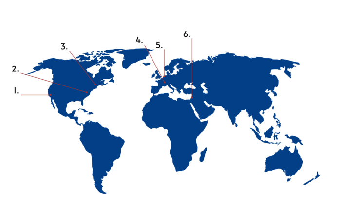
| Foreign partner (unit name) | Principal investigator(s) | Area of cooperation | |
|---|---|---|---|
| 1. | CHLA, Los Angeles, CA | Robert Shaddy | ABM grant on DMD/BMD |
| 2. | National Heart, Lung and Blood Institute, Bethesda, USA | Peter Kellman | Quantitative perfusion mapping in microvascular disease models, using a dual sequence prototype |
| 3. | Harvard University, Boston, MA University of Groningen | Maaike van den Boomen, Ronald Borra | Myocardial microcirulation reactivity assessment with a prototype SMS-GESE-EPI T2/T2 mapping |
| 4. | University Hospital, Lausanne | Ruud van Heeswijk | 3D T2 myocardial mapping prototype sequence – assessment in focal myocardial inflammation |
| 5. | Hart Long Centrum Leiden | Dr. V. Delgado | Bicuspid aortic valve – left ventricular strain evaluation as prognostic tool |
| 6. | Product Manager, ANGIO Mentor, Healthcare, ISRAEL | Shani Fargun | The goal of the current program is to assess the new “See the Invisible” AR add-on to the ANGIO Suite medical simulator. |

Key publications
1. Clinical Features and Outcomes of Takotsubo (Stress) Cardiomyopathy. N Engl J Med. 2015 Sep 3;373(10):929-38. doi: 10.1056/NEJMoa1406761.PMID: 26332547
2. Prognostic implications of left ventricular global longitudinal strain in patients with bicuspid aortic valve disease and preserved left ventricular ejection fraction. Eur Heart J Cardiovasc Imaging. 2020 Jul 1;21(7):759-767. doi: 10.1093/ehjci/jez252.PMID: 31633159
3. Influence of observer-dependency on left ventricular hypertrabeculation mass measurement and its relationship with left ventricular volume and ejection fraction: comparison between manual and semiautomatic CMR image analysis methods. PLoS ONE 2020 15(3): e0230134.
4. Brain resting state functional magnetic resonance imaging in patients with takotsubo cardiomyopathy an inseparable pair of brain and heart. Int J Cardiol. 2016 Dec 1;224:376-381. doi: 10.1016/j.ijcard.2016.09.067. Epub 2016 Sep 18.PMID: 27673694
5. Left ventricular volumes and function affected by myocardial fibrosis in patients with Duchenne and Becker muscular dystrophies: a preliminary magnetic resonance study. Kardiol. Pol. 2020 : t. 78, nr 4, s. 331-334, bibliogr. 14 poz.DOI: 10.33963/KP.15223 (IF= 1.874)
6. Required temporal resolution for accurate thoracic aortic pulse wave velocity measurements by phase-contrast magnetic resonance imaging and comparison with clinical standard applanation tonometry. BMC Cardiovasc Disord. 2016;16(1). doi:10.1186/s12872-016-0292-5